Murder, she wrote

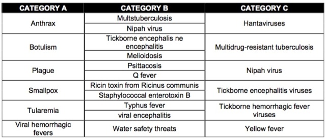
Canterbury, 1999. A woman was brutally murdered and apparently no evidences were left at the crime scene that could help the police to solve the case. However, as TV shows and black novels have taught us, a small sample of tissue, a drop of blood, saliva or hair may be sufficient to exonerate the innocent, identify suspects and convict the guilty. The golden clue to catch her killer, as a forensic scientist discovered, is under the nails of the woman. There is a tiny trace of murderer’s skin under her fingernails when in a desperate final attempt to preserve her life she scratched her assailant. It wasn’t a perfect crime and the murderer had left behind an important clue after all, their DNA.

DNA is a molecule that contains the hereditary material that makes a person unique and unrepeatable. DNA is a polynucleotide molecule composed of two antiparallel strands twisted into a double helix structure, which is held together by hydrogen bonds between the paired bases. Nucleotides are known as the building blocks of DNA. There are four basic nucleotides in a DNA chain linked by covalent bonds: adenine (A), guanine

(G), cytosine (C) and thymine (T). It is the sequence of nucleotides on a DNA strand what determines individual hereditary characteristics. Knowing that the human genome is 3.000 million base pairs in length and the genetic differences between individuals are only 0.1%, the identification of a person analyzing their DNA sounds like looking for a needle in a hayrack. Even though 99.9% of the DNA is identical across all humans, the small percentage of DNA that differs is enough to identify individuals, except for monozygotic twins who have identical DNA profiles. In 1984, Alec Jeffreys developed the genetic fingerprinting, a method used to identify individuals using DNA samples. Although the first DNA test was carried out to solve an immigration case, it soon became clear the potential of DNA profiling as a powerful tool to fight against crime.


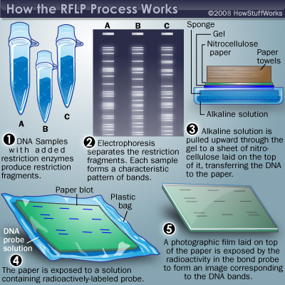
The technique of genetic fingerprinting is based on the study of highly variable DNA regions (or polymorphic) that are unique to almost every individual. The first procedure used for DNA fingerprinting is known as Restriction Fragment Length Polymorphism (RFLP ). In this method, restriction enzymes are used to cut DNA at a specific nucleotide sequence producing numerous DNA fragments of different length. The resultant DNA fragments are loaded onto a gel and separated according to their size using an analytical technique called electrophoresis. An electric field is applied across the gel and the fragments migrate toward the positive electrode, the larger molecules will move more slowly than smaller molecules that will travel farther through the gel. Then, the gel is processed further using a blotting technique. The double-stranded DNA is denatured into single strands and transferred to a nylon membrane. The membrane is incubated with labeled DNA probes. DNA probes are designed single-stranded DNA fragments complementary to the fragment of interest. Therefore they will detect its complementary sequence, excluding all the other fragments. The segment attached to the probe can be visualised by exposure to X-ray film where they form a pattern of dark marks that can be analysed.

The main inconvenience of analyzing DNA using the RFLP method is that it requires large amounts of high-quality DNA, which are hardly found in a crime scene. More recent approaches of this technique involve the use of the Polymerase Chain Reaction (PCR) that allows the amplification of smaller DNA fragments within hours. Nowadays, the use of Short Tandem Repeats (STR) rather than Variable Number Tandem Repeat (VNTR are used in the original procedure) is widespread as they can be used to analyse degrade DNA samples.

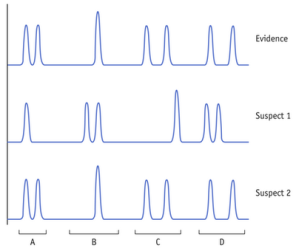
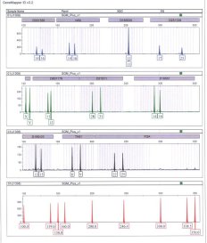
If the DNA profile from a suspect matches the DNA evidence found in a crime scene, it is highly probable that the suspect is linked to the crime scene (although it may happen, it is unlikely that two DNA profiles match by chance)


DNA profiling has also many other applications such as:

  • Parental testing
  • Wildlife management
  • Medical diagnosis
  • Ancient DNA
  • Immigration disputes


In 1995, UK was the first country in the world to set up a DNA database (NDNAD), which hold DNA profiles used to identify suspects of crimes. It not only contains DNA profiles from offenders, but also from individuals who have been arrested but not convicted. Although it has been recognized as an important tool in criminal investigation, it also has raised numerous concerns. DNA contains private information about ethnicity, health or genetic relationships. Therefore, if this information is misused privacy will be compromised

Posted in Uncategorized | Leave a comment

The genetic revolution is here

2015 is gone and it is time to look back and take stock of all what we have achieved throughout the year and make new promises for the coming year. 2015 was a year of many changes all over the world and overall it has also been a great year in the field of science and technology. Here, there is a short list of some of the most important scientific discoveries made in 2015:


A vast majority of the scientific community has named CRISPR-Cas9 as the most important scientific discovery of 2015. CRISPR-Cas9 stands for Clustered Regularly Interspaced Short Palindromic Repeats and is known as the immune system of bacteria and Achaea.

The vertebrate immune system is a complex network of specialized molecules, cells, tissues and organs that are responsible for defending the body against foreign substances. Other animals, such as insects, molluscs or crustaceans also have immune system, although it is much simpler. But how can unicellular organisms like bacteria have an immune system?

Viruses can infect bacteria, these viruses are known as bacteriophage. In a simple way, the infection cycle starts when the virus binds to the surface of the bacteria in a specific place and injects its nucleic acid into the cell. Once inside, the viral DNA takes control of the cellular machinery to facilitate the replication of the viral genetic material and the synthesis of viral proteins. Finally, the new virions are released into the environment so they can infect the adjacent cells.

In order to protect themselves from foreign DNA, bacteria have their own defense mechanisms, the restriction-modification systems. The system consists of two enzymes, methylases that add a methyl group (CH3-) to DNA at a specific site, and restriction enzymes that recognize a specific nucleotide sequence and cut the DNA near or at that particular place (restriction site). Bacteria have the ability to methylate their DNA, which allows them to distinguish between foreign DNA and their own DNA. The restriction enzymes cannot cut the methylated DNA, so the foreign DNA would be recognized because it is not methylated.


There is also another way to detect and destroy viral DNA: the CRISPR-Cas system.

Within the bacterial genome there are some regions where the nucleotide sequences are palindromes, that is to say, sequences that read the same backward and forward (“A man, a plan, a canal- Panama!”). In the bacterial genome, the alternation between palindromic DNA repeats and spacers sequences (short segments of foreign genetic material) is present in a particular region of the genome known as CRISPR locus. CRISPR regions are associated to genes that encode particular nucleases; Cas (CRISPR-associated sequences), which are proteins specialized for cutting DNA. When a virus attacks bacteria and injects its DNA, the presence of viral DNA provokes the activation of the CRISPR system. The CRISPR sequences are transcribed into RNA to target and cleave a specific site of the viral DNA at two strands. The CRISPR-Cas complex allows to the Cas enzymes to cut the foreign DNA, so the ARN works as a guide to the Cas enzyme, which acts as a scissor, it cuts and inactivates the viral DNA.


Emmanuelle Charpentier and Jennifer Doudna are worldwide known for demonstrating that it was possible to create “guide” RNA that can target any DNA region of any specie and cut it by using a particular enzyme, Cas9. What is more, it also allows the introduction of specific sequences, so the genome can be edited.

The CRISPR-Cas9 technology marks the beginning of a new era in genetic engineering in which you can edit, correct and alter the genome of any cell in an easy, quick, cheap and accurate way. In the future it will serve to cure diseases whose genetic cause is known but they are currently incurable. For example, the MIT (Massachusetts Institute of Technology) announced in March 2014 they had reversed an adult mouse liver disorder (type I tyrosinemia) using this genetic technology.

The ethical controversies of using the CRISPR system are related to the process of modifying the human germ line. In an article published in April 2015, Chinese scientists described the use of CRISPR system to edit the genome of human embryos that would create a heritable modification, which would affect future generations. Although the embryos were nonviable it initiated an ethical debate about the use of this editing tool.

Although this technique still requires many improvements for its effective application, there is no doubt that CRISPR-Cas9 has much to offer and surely we will hear a lot more about it in the future. As John Travis, editor of Science News said: “For better of worse, we all now live in CRISPR’s world”.

Posted in Gene editing | Leave a comment

The power of three

On 3 February 2015, The House of Lords gave the green light to a new and controversial form of in vitro fertilization, the mitochondrial donation. The treatment involves the intervention in the fertilization process in order to remove defective mitochondria from the mother and replace them with healthy mitochondria from a donor’s egg to prevent the transmission of mitochondrial diseases to the offspring, thus giving rise to the so-called three-parent babies. UK became the first country in the world to allow the mitochondrial donation. But before going in further details, I will talk briefly about mitochondria and mitochondrial diseases.

Mitochondria are double-membrane organelles present in most eukaryotic cells. One of the most important functions of mitochondria is the production of ATP, which is essential in most of the cellular processes. The two mitochondrial membranes create two different regions in the mitochondria, the matrix and the intermembrane space. The inner membrane separates the mitochondrial matrix from the intermembrane space, which is the region between the inner and outer membrane. The mitochondrial matrix contains ribosomes and a small amount of mitochondrial deoxyribonucleic acid (mtDNA). Mitochondrial DNA is circular; it has 16,569 DNA base pairs and contains 37 genes that encode 13 proteins, 22 tRNAs and 2 rRNAs.


Mitochondrial diseases are a group of inherited chronic disorders resulting from a deficiency of one or more proteins localized in mitochondria and which are involved in the metabolism. Mitochondrial disorders may be caused by mutations in mitochondrial or in nuclear DNA due to mutations in genes coding for proteins involved in the correct functioning of the mitochondria. Although within the cell, mitochondrial DNA represents a very low percentage of genetic information, the severity and symptomatology of diseases is very extensive. The most affected organs and tissues are: brain, muscle, eye, ear, heart, endocrine system, liver, kidney, intestine, skin, bone marrow and blood. Therefore the range of symptoms is very wide from dementia to poor vision and neuropathic pain. Some of these diseases are:

  • Leber’s Hereditary Optic Neuropathy (LHON)
  • Kearns-Sayre syndrome (KSS)
  • Leigh’s Disease
  • Pearson syndrome
  • Progressive external ophthalmoplegia (PEO)


So, if the problem is faulty mitochondrial DNA, let’s remove it!

The most developed techniques to prevent the transmission of mitochondrial diseases from mothers to children are: maternal spindle transfer (MST) and pro-nuclear transfer (PNT). Both of them are based on IVF and both require 3 people:

  • The father, whose sperm fertilizes the egg.
  • The mother, whose mitochondrial DNA has been eliminated only giving to the new embryo the DNA from the nucleus of the egg.
  • The female donor, who gives to the new embryo their mitochondrial DNA.

In the first technique, Maternal Spindle Transfer (MTS), two eggs are required, one from the mother and one from the donor. The nucleus, which contains most of the genetic material, is removed from the eggs. The donor nucleus is destroyed and replaced by the mother nucleus. Therefore, the resulting egg contains nuclear DNA from the mother and healthy mitochondria from the donor. Finally, the egg is ready to be fertilized by the father.

In the second technique, Pro-nuclear Transfer (PNT), both eggs, from the mother and from the donor, are fertilized with the father’s sperm. The donor pronuclei are removed and destroyed and the parent’s pronuclei are inserted into the donor’s egg. The new embryo cell contains both, nuclear DNA from the parents and healthy mitochondria from the female donor. Once ready, the embryo will be transferred to the mother’s uterus.


There is no doubt that an endless number of concerns related to ethical, legal and social issues have arisen due to this novel method. However, we should bear on mind that mitochondrial donation could mark an end to certain diseases and it also gives hope to many families with genetic predisposition to suffer serious and incurable diseases.

Posted in IVF | Leave a comment

The Barcode of Life

FFDL6YBDKD

Throughout history scientists have identified around 1.7 million species with which we share the planet. However, it has been estimated that there are over 3-30 million unknown species on Earth. Therefore, it exists a huge commitment within the scientific community to study and inventory all this biodiversity.

Traditionally, specimens are identified and classified according to a set of universal rules that also establishes evolutionary linkages and kinship among different organisms. Taxonomy is the branch of science responsible for this systematic classification. This classificatory system is a slow and thorough process that requires specialists with high identification skills. In many occasions, taxonomists, as these specialists are known, have many difficulties to access to specialized literature because of the language, format or geographical issues, which undermines the classification process.

What is Taxonomy?

Due to these and other impediments, in 2003 Dr. Paul Hebert proposed the identification of living organisms by using standardized genomic regions as if they were barcodes. The DNA barcoding is based on the same functional idea of scanners in supermarkets that distinguish commercial products using the Universal Product Code. Instead, this technique uses a short DNA sequence to identify species. But, which region of the genome is a suitable barcode? The optimal DNA barcode needs to:

  • Be present in all organisms.
  • Distinguish all species.
  • Be easy to amplify and sequence.


A small fragment of 648 base pairs in the mitochondrial gene cytochrome c oxidase subunit 1 (COI) was selected as the standard barcode for animals. However, COI is a poor barcoding gene for plants and fungi. Plants exhibit insufficient mitochondrial sequence variation therefore two chloroplast genes matK and rbcL have been established as core-barcode for plants. On the other hand, COI region in fungi cannot be easily amplified and not all the fungal species have mitochondria, therefore one nuclear gene region, the internal transcription spacer (ITS), has been chosen as the universal fungal barcode.

The methodology of the DNA barcoding is “quite simple”. When a specimen is collected, its DNA is extracted from a tissue sample. From the sample taken, the PCR (Polymerase Chain Reaction) technique is used to obtain numerous copies of the DNA barcode region. Then the amplified gene is sequenced and represented by a succession of letters CATG (corresponding to the different nucleotides: cytosine, adenine, thymine and guanine). The DNA sequence is uploaded to a database where it can be identified if it matches with species that are already present in the database. It may also occur that the DNA sequence does not match with any of the species therefore the novel DNA sequence might suggest that the database is incomplete and the unknown sequence corresponds to possible new specie.

DNA barcodes can be used in many different ways, such as:

  • Pest control.
  • Identification of threatened, endangered and invasive species.
  • Control the illegal wildlife trade.
  • Differentiate males and females of each species.
  • Identification of species at any stage of life.
  • Food traceability (a tool to ensure food safety and quality).
  • Identification of disease vectors.

The DNA barcoding technology was introduced as a new technique for specimen identification and species discovery, which can complement the work done by taxonomists. Despite having its limitations, experts are working on improving mistakes to achieve the standardization and reliability of the technique.

If you want to know more about how taxonomists work, how the DNA barcoding technique is used to assess biodiversity and many other interesting things I strongly recommend a visit to the Natural History Museum in London. I really enjoyed my visit and I had the chance to talk with experts and understand their work. It is well worth a visit specially during this Christmas break.

Posted in DNA Barcode | Tagged | Leave a comment

“All is fair in love and war”

Human beings are extraordinary. All the accomplishments made by human throughout history have demonstrated the unlimited power and potential of our mind. However, our past has also showed the dark side of human nature.

Biological weapons are biological agents deliberately spread in order to cause death or incapacitation among humans, animals and plants. The main biological agents used as weapons are:

  • Bacteria: unicellular organisms that grow and reproduce very
    rapidly.
  • Viruses: entities that need a host cell in which to live and reproduce.
  • Toxins: substances produced by numerous organisms that can be highly poisonous.

Diseases caused by biological agents can be classified into different categories according to how easily the disease can be spread, how severe the public risk is and how lethal the disease can be. Thus, there are three categories A, B and C. Category A includes high-priority agents whereas category C includes the lowest-priority as well as emerging pathogens.

foto

Classification of the diseases into three categories

A biological attack represents an increasing threat to public health mainly because the agents used are odorless, tasteless, invisible and therefore they can be spread silently in a wide range of ways (in aerosols, explosives, food or water and transmitted from person to person). Furthermore, biological weapons are relatively easy and cheap to make but difficult to detect. Only a small amount is needed to cause thousands of deaths or an environmental catastrophe.

The first uses of living organisms as a weapon are dated several centuries ago. The use of plagues, epidemics and diseases in the wars has been in the mind of man well before the scientific development. During WWI and WWII governments led experiments with biological and chemical weapons, some of them were even used to attack the civilian population.

In 1972, as a consequence of the impact of biological weapons on humanity, over 103 nations signed an agreement in which the development, production and stockpiling of biological and toxin weapons were prohibited. However, this treaty was ineffective and many bioweapons were developed illegally. Moreover, technological advances in genetic engineering marked a turning point in the use and development of bioweapons, increasing the risk of biowarfare. By using genetic engineering, biological agents can be enhanced and produced in larger amounts. They can also be genetically modified not only to be more toxic and infectious but also to transform harmless organisms into lethal organisms by introducing genes from a highly pathogenic organism.

Nowadays, biological weapons are among the biggest threats to human existence. One of the most remarkable concerns is that the genome of microorganism that cause tuberculosis, cholera, leprosy, anthrax and plague is accessible on the Internet making easy the possibility of designing a biological weapon quickly and cheaply.

We are aware that science has discovered many things that can contribute to man’s adaptation to nature; however, this adaptation depends on the correct application of science.

Posted in Biowarfare | Leave a comment

Protozoa, Plasmodium and Paludisme.

Protozoa are unicellular eukaryotic microorganisms. Although they are difficult to classify due to their abundance and diversity (more than 50,000 have been described) the most common classification is according to their mechanism of motility. Therefore, there are four subcategories: amoebae, flagellates, ciliates and sporozoan. Protozoa are also defined as ubiquitous, that is to say, they can be found in almost every kind of environment, mainly in moist and watery habitats. They can live as free entities or in association with other species. This symbiotic relationship can be commensalism, mutualism or parasitism.


Plasmodium is a genus of parasitic protozoans of the sporozoa subclass coccidian. Among all plasmodium species, there are four that can infect humans: P.falciparum, P.vivax, P.ovale and P.malariae, each of which displays different clinical symptoms.

Paludisme, most commonly known as Malaria due to the variety P.malariae, is an endemic disease caused by plasmodium. Malaria is transmitted by Anopheles mosquitoes, which carry plasmodium on their salivary glands. Only female mosquitoes can infect humans, as they require nutrients from blood to nourish their eggs (male mosquitoes are exclusively nectar feeders).

The infection starts when a mosquito bites a human and injects its saliva along with plasmodium (in the form of sporozoites). Parasites travel through the bloodstream and reach the liver and penetrate liver cells (hepatocytes). Inside the liver cells, sporozoites multiply via asexual replication and the daughter cells, called merozoides, break the liver cells and re-enter the circulatory system. In the next stage, merozoides invade red blood cells (erythrocytes) and multiply asexually again until the infected cells burst and the merozoites are released to the bloodstream. At this point, merozoites can either infect new erythrocytes and restart this process or develop into gametocytes (sexual form of the parasite).

But the cycle does not end here. When a mosquito bites a human who has previously been infected, the gametocytes are transferred to the mosquito and the formation of gametes or gametogenesis occurs. Male gametes fertilise female gametes, they fuse together and form zygotes, which will develop in ookinetes. The ookinetes are motile and migrate to the gut epithelial cells and form oocysts that will reproduce asexually to finally produce sporozoites. Then, the sporozoites migrate to the mosquito salivary glands so they can infect a new human.

Malaria is one of the most common infectious diseases in poor tropical and subtropical countries of the world. According to The World Malaria Report 2014 around 3.3 billion people are at risk of malaria and 1.2 billion are at high risk. Moreover, malaria causes an estimated 584.000 deaths every year. Currently, there is no effective vaccine for malaria.

The media has been long used as a tool to influence the public perception on many issues. Many films have been released to educate on malaria. One that I found particularly curious is The Winged Scourge, an animated short produced by The Walt Disney Studios, which defines the anopheles mosquito as the public enemy no. 1. This animated propaganda also highlight that the best way to fight Malaria is by fighting mosquitoes.

(Please note that this short was released in 1943. The methods used to prevent and control malaria such as spraying oil on water and the use of gas Paris Green  are no longer in use)

Posted in Malaria | Leave a comment

The key to immortality

We have heard a lot about 3D printing over the last few years, however, this emerging technique is not as new as we may think.

The first approach to 3D printing was in 1860 when the French artist François Willème developed the “photo-sculpture”. Very briefly, Willème photographed an object, which was surrounded by 24 cameras (arranged every 15 degrees), and with all the photos taken he created the 3D representation of the object. The revolution came in the 1980´s and it was led by Chuck Hull, an American engineer who invented the “stereolithography” which allows the creation of complex three-dimensional objects from CAD drawing (computer-aided design) within hours.

Ever since then, 3D printing has experienced an unexpected expansion and nowadays it has endless applications. By using 3D printers it is possible to make a wide range of products, from guns to parts for vehicles. Scientists also found that printers could be useful resources in the medical field and soon they started to produce dental implants and prosthetics using different materials like plastic or ceramic. One of the advantages of this method is that it allows to print highly customised body parts, reproducing accurately the specific needs of each patient. However, human beings are ambitious and we now face a new challenge, printed tissues and organs.

Even though printing complex organs is still a challenge, printed human tissue is already a fact.

Bioprinting is the technique used to print human tissue from living cells using a 3D printer. As any other printer, bioprinters need ink to start the process. The “bioink” used is designed according the patient’s specifications and it is constituted by their own cells, either from a biopsy or stem cells. Once the cells multiply in a growth medium, they are loaded into cartridges and the bioprinting process starts. Another cartridge is also loaded with hydrogel which provides the right conditions for the cells to survive and it also acts as a space holder. This step (hydrogel/cells layer) will be repeated as many times as required and during this process, the cells will fuse together naturally. The last stage is a maturation and growing phase that may take several weeks, during this time the hydrogel is removed.

There are few developments done in the science field that exist without ethical controversies and certainly brioprinting raises a large number of ethical questions.

  • Who would have access to treatments?
  • How wait lists will be managed?
  • Is it safe?
  • Could be an organ repaired, rebuilt or replaced forever?
  • Could it be used for human enhancement?

So far, printed tissues are used in a small-scale  for medical research, drug discovery and toxicity testing. The next step would be the design and creation of simple tissues which can be implanted into organs. However, the last and most challenging purpose of bioprinting technology is to be capable of printing an entire human organ.

Although there is still a long way, bioprinting has the potential to become a revolutionary technology.

Sci-fi or real life?

Posted in Bioprinting | Leave a comment